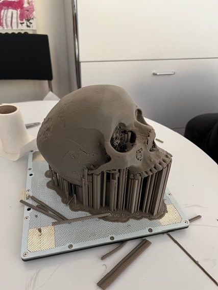
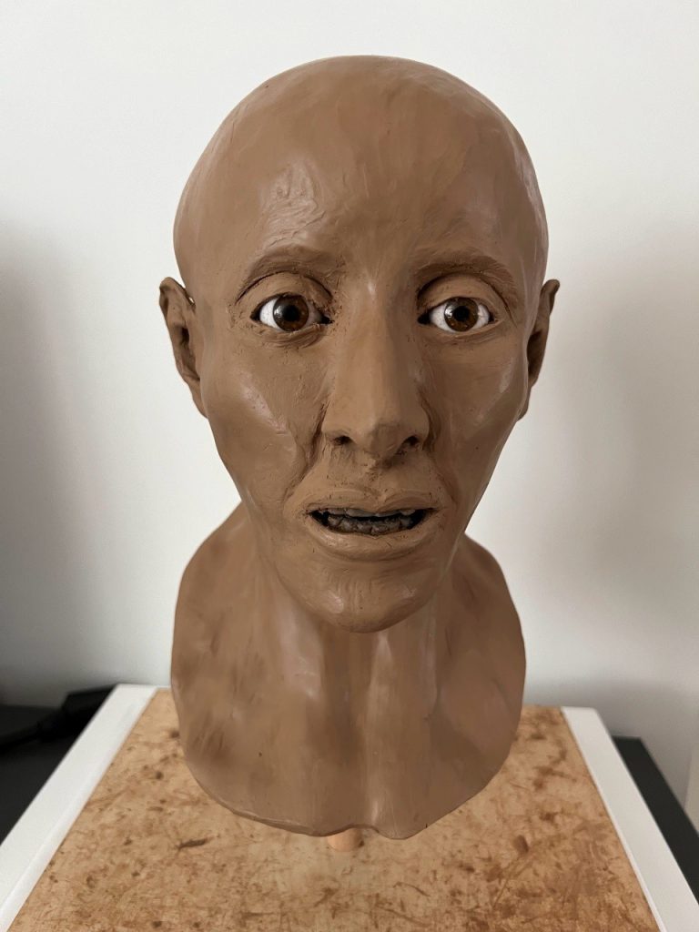
From the computed tomography scan realized at the IEM, a facial reconstruction was elaborated by Dana Ackermann in 2022. The work was done within the framework of a Maturitätsarbeit at the Kantonsschule Limmattal under the supervision Dr. Anna-Katherina Holenweg. The practical reconstruction work was supervised by Dr. med. Patrick Eppenberger at the IEM. If not specified, the images were taken from Achermann’s Maturitätsarbeit and copyright of the Kantonsschule Limmattal. Ackermann used the Gerasimov method (Manchester Method), where muscle and skins are gradually built up by hand over the 3D printed scull. Skin thickness is based on specific data on ethnicity and indicated with tubular spacers that are placed all over the scull. Age, sex, way of life and nutrition are all impact the thickness of muscles, glands and fatty tissues.
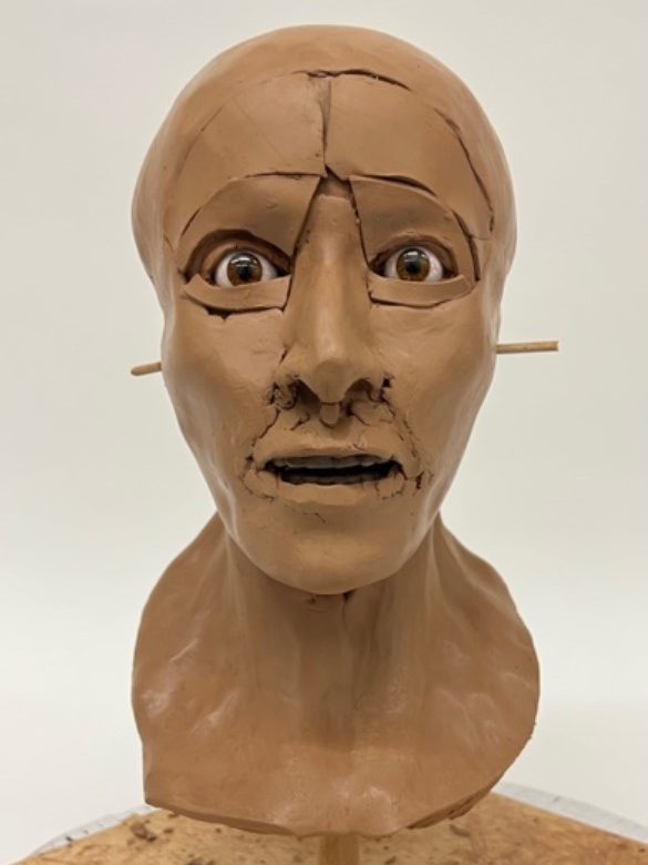
Once the spacers in place, every muscle and gland is carefully modelled by hand. Symmetry, size and form of the nose is reconstructed according to ethnicity, but also on the shape and size of the apertura piriformis, the pear-shaped nasal cavity of the scull. The dental record informs the shape and size of mouth and lips. The ears are the most difficult and least accurate parts, as the scull does not inform us on how they looked like. They are modelled according to available data on ethnicity. The size of the inset glass eyes is chosen according to the aperture of the eye sockets. Once all muscles and glands have been placed, the skin is laid over them in several pieces and joins are smoothed over.
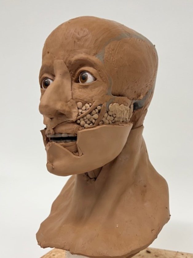
The degree of finish is the one that is usually used in forensic investigations in order to visualize and identify human remains. At this stage modelling marks are still visible; the skin tone is the one of the modelling material, and no attempt is made for the reconstruction of facial hair. This said, it gives us a very real first impression of what Ta-Sherit-en-Imen looked like some 2800 years ago. The careful and excellent work of Dana Ackermann can only be applauded, more particularly as this was her first attempt of a facial reconstruction.
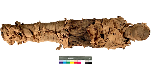
In the late 19th century, the mummy of Ta-Sherit-en-Imen was partially unwrapped, exposing the resin coated bandages of the abdominal area. This was most likely done in the search for amulets or to access the mummified body. On the right hip some of the bandages were removed exposing the parchment-like skin. Fortunately, the inner layers were heavily impregnated with resinous matter and prevented any further desecration of the body. The restoration of the mummy’s bandages was performed by Laura Flückiger as part of her MA thesis (https://sonar.ch/hesso/documents/321908) under the supervision of conservators Valentin Boissonnas (HE-Arc), Mimi Levèque (Massachusetts, USA), and Agniezka Jucker-Woos (Abegg Stiftung Riggisberg).
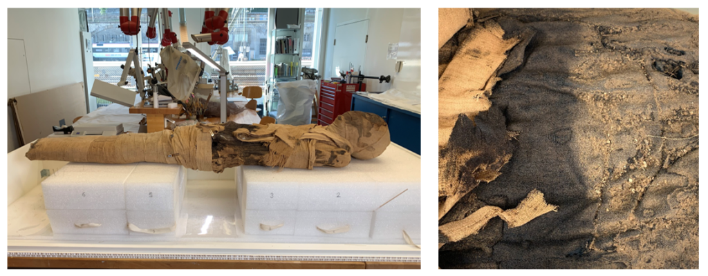
The conservation project aimed at characterizing the different linen bandages, understanding their sequence, locating the position of loose bandages, and placing as much of them as possible into their original position. The conservation work was done in a designated conservation lab at the HE-Arc that could not be viewed or accessed by the general public. The fragility of the mummy prohibited turning the body during treatment. A polyethylene foam support was constructed in a series of sections that allowed to temporarily access any section of the mummy while the rest of the body remained well supported.
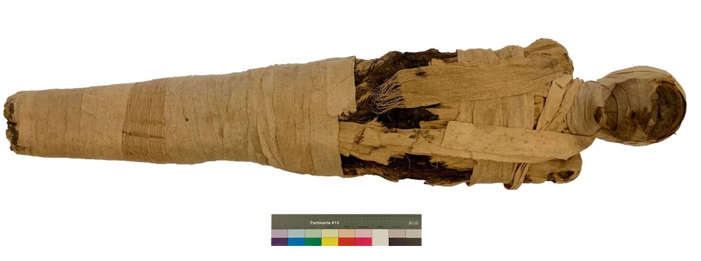
The mummy on its temporary working support and during cleaning of surface dust and debris
All textile bands were first carefully cleaned of dust and particles that had accumulated over the years of uncontrolled storage. The presence of mold on some of the textiles necessitated the use of protective gear and the use of HEPA filters for the low suction hoover. The removal of dust and debris was done with fine brushes and tweezers. Many bandages presented creases and folds that had formed after their unwrapping. These were reshaped by controlled humidification using humidified blotting paper over a semi-permeable Sympatex layer placed over the textile and sandwiched between two sheets of Melinex.
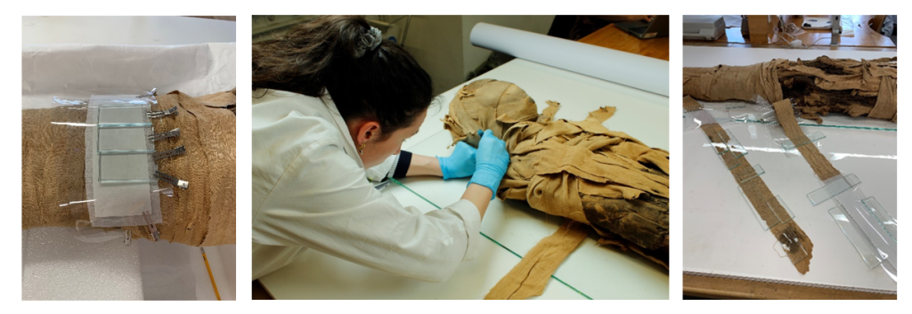
Reshaping and repositionning of the bandages after cleaning
Torn bandages and areas of loss were stabilized by sowing them into two layers of very fine tinted nylon netting. To keep all bandages in place and avoid future damage to fringes and loose threads, a fine tinted nylon gauze was placed over the repositioned bandages. Stitching was done within the nylon mesh. A few loose threads and textile areas that could not be secured mechanically by netting were secured with a 4% methylcellulose in deionized water. Bandages that could not be attributed to a specific position were kept apart as research material.
Conserving the mummy of Ta-Sherit-en-Imen was a rare privilege and a can be considered a continuation of the work that embalmers began in ancient Egypt. It gave back dignity to the deceased and enabled her to continue her journey to the beyond and be reborn over and over again. The treatment methodology for Ta-Serit-en-Imen was done with a maximum respect for her person and in accordance today’s conservation ethics. The trauma from the opening of the bandages, at a time when mummies were just another commodity, remains visible and has become an integral part of Ta-Sherit-en-Imen’s history.